
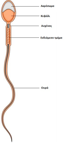
Spermatozoon anatomy and physiology
The typical spermatozoon is about 50 μm long and consists of a head and a tail. The head is oval in shape and is flattened with a length of 4-5 μm and a width of 2.5-3.5 μm and with a ratio of length: width 1.50 to 1.75. The nucleus of the spermatozoon occupies about 65% of the head and consists of densely packed chromosomal material (mainly DNA) and basic proteins. The anterior part of the head is covered by a sac-like structure, which is called the acrosome. This contains the necessary enzymes for the sperm to penetrate the various layers surrounding the ovum and its transparent zone.
The tail is about ten times the length of the head. It is located within a thin shell and has two central paired microtubules surrounded by 9 pairs of circumferential fibers from the neck to the end of the tail. The intermediate part is the anterior part of the tail and consists of the densely arranged mitochondria of the spermatozoon. The connection between the tail and the intermediate part is called the terminal ring. Overall, the microtubules, receiving energy from the mitochondria in the intermediate part, are the organ that enables the sperm to move.
Sperm motility is the most important factor in transporting sperm through the cervix, although other factors may play a role. In the middle of the menstrual cycle, when the quality of cervical mucus is optimal, sperm enter the cervix rapidly (within 30-60 seconds after ejaculation). Some spermatozoa enter the uterus and reach the fallopian tubes very quickly. Others are "stored" in the cervical mucus and are gradually released for up to 2 to 4 days. The presence of sperm in the cervical mucus can be observed even 7 days after intercourse.
Of the millions of sperm, which are normally present in the semen, only 0.1-1% reach the uterus. Finally, it’s only about 1000 - 5000 that can reach the fallopian tubes, where fertilization takes place. All excess spermatozoa are removed by the process of phagocytosis, from the white blood cells that reach the uterus in large numbers within 10 to 24 hours after the entry of the first spermatozoon. Some sperm can also escape from the open end of the fallopian tube and be found in the peritoneal cavity.
The first spermatozoon reaches the fallopian tube within 5 - 15 minutes after intercourse, while the typical time for most spermatozoa is from 2 to 24 hours. However, although spermatozoa are fully mature as cells, they cannot fertilize the egg, until certain morphological and biochemical changes occur. Spermatozoa undergo these changes in the cervix, uterine cavity, and fallopian tubes.
